Clinical Science Review · Vol. 3 · For healthcare professionals
Androstenedione, Testosterone, and DHT
Abstract
- Rationale
- Androgen status is often reduced to a single testosterone value. But testosterone sits in the middle of a metabolic sequence: a weak precursor, androstenedione, lies upstream, and the most potent androgen, dihydrotestosterone, lies downstream. The same testosterone concentration can arise from — and give rise to — very different states.
- Key distinctions
- Androstenedione is a weak precursor androgen of adrenal and gonadal origin; testosterone is the principal circulating androgen; DHT is the most potent, a high-affinity non-aromatisable androgen-receptor ligand generated in target tissues by 5α-reductase. Because each conversion is enzymatic — 17β-HSD, 5α-reductase, aromatase — the ratios between the three analytes encode enzyme activity and androgen source.
- Clinical implications
- Measuring all three and reading their ratios localises androgen excess (adrenal versus ovarian or gonadal), exposes 5α-reductase and aromatase activity, and makes the predictable metabolic patterns imposed by 5α-reductase inhibitors and aromatase inhibitors legible.
- Conclusion
- A single androgen is a point; the pathway is the picture. Panel measurement with the ratios yields information no single analyte provides — provided the assay is accurate across the relevant ranges.
Keywords:androstenedione · testosterone · dihydrotestosterone · 5α-reductase · aromatase · 17β-HSD · T:A4 and DHT:T ratios · hyperandrogenism · LC-MS/MS
1. Background
Androgen assessment in routine practice is commonly a single testosterone measurement. Yet testosterone is a midpoint in a metabolic sequence: a weak precursor feeds it, a more potent product derives from it, and a parallel route converts both to estrogens.
Reading only the midpoint discards where the androgen came from and what it becomes.
This review makes the case for measuring the three principal androgens together — androstenedione, testosterone and DHT — and interpreting the ratios that encode the pathway and its enzymes.
2. One pathway, three androgens
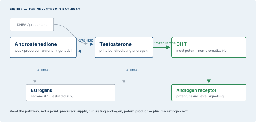
Androstenedione, a weak androgen produced by adrenal and gonadal tissue, is converted to testosterone by 17β-hydroxysteroid dehydrogenase; testosterone is reduced to dihydrotestosterone by 5α-reductase in target tissues; and both can exit the androgen pathway to estrogens via aromatase.
DHT is the most potent androgen — the highest-affinity androgen-receptor ligand, and non-aromatisable — and much of its action is intracrine, generated within skin, hair follicle and prostate.3,10
The corollary is that the three analytes report different things:
- Androstenedione reports precursor supply
- Testosterone reports circulating androgen
- DHT reports potent, tissue-level androgen activation
3. The ratios encode enzyme activity and source
Because each step is catalysed by a distinct enzyme, the ratios between the three analytes are themselves informative:
- T:A4 reflects 17β-HSD conversion of precursor to active androgen
- DHT:T reflects 5α-reductase activity
- A disproportionately high androstenedione points upstream — toward an adrenal or ovarian source, or an enzymatic block
In polycystic ovary syndrome, simultaneous LC-MS/MS profiling shows elevated testosterone and androstenedione together with an altered product-to-precursor ratio, localising the androgen excess to the ovary rather than the adrenal.2
A single analyte cannot separate these possibilities; the pathway can.
4. Metabolic patterns imposed by enzyme inhibitors
Two common therapies rewrite the pathway in predictable, measurable ways.
5α-reductase inhibitors (finasteride, dutasteride) block the conversion of testosterone to DHT: serum and scalp DHT fall — dutasteride lowering DHT more completely than finasteride, dose-dependently — while testosterone rises, inverting the DHT:T ratio.4 Finasteride reduces circulating DHT by roughly 70%, and prostatic DHT still further.5
Aromatase inhibitors block the conversion of testosterone to estradiol, and androstenedione to estrone: estrogens fall and androgens accumulate, so testosterone rises as its estrogen exit is closed.7,8
In each case it is measurement of the pathway — not of a single analyte — that makes the shift legible.
5. Applications favouring measurement of all three
5.1 Hyperandrogenism and PCOS
Total and free testosterone are the first-line laboratory tests for biochemical hyperandrogenism, with androstenedione and DHEA-S adding source information when testosterone is not elevated; the 2023 International PCOS Guideline analysis supports this tiered approach and recommends LC-MS/MS for accuracy.1 Adding androstenedione and reading the product-to-precursor ratio helps distinguish ovarian from adrenal excess.2
5.2 Congenital adrenal hyperplasia
In 21-hydroxylase deficiency, androstenedione — together with 17-hydroxyprogesterone — is a core marker of androgen control, monitored alongside testosterone.6
5.3 Androgen-dependent skin and hair conditions
DHT and 5α-reductase drive androgenetic alopecia, hirsutism and acne.3 Measuring DHT and the DHT:T ratio characterises peripheral androgen activation.
5.4 Therapy and disease monitoring
The panel tracks the effects of 5α-reductase and aromatase inhibitors, and in prostate cancer it exposes what a single value hides: under androgen deprivation, DHT can persist through intracrine and backdoor synthesis, and androstenedione becomes associated with testosterone in castration-resistant disease — patterns visible only when precursors and products are measured together.9,10
6. Analytical considerations
Androgens span a wide dynamic range and are frequently low — in women, in children, and under pharmacologic suppression — where direct immunoassays are unreliable, particularly for androstenedione and DHT; radioimmunoassay overestimates androstenedione relative to mass spectrometry.2
The 2023 International PCOS Guideline analysis explicitly recommends LC-MS/MS for androgen measurement given its accuracy, and validated panels quantify androstenedione, testosterone and DHT from a single specimen.1
Where the DHT:T ratio can and cannot be computed
The ratio arguments above depend on a quantifiable DHT, and that is a real constraint rather than a theoretical one.
Clinicore reports DHT from 10.125 ng/dL. That covers most of the female reference interval and the elevated concentrations characterising androgen excess — hirsutism, PCOS, androgenetic alopecia, suspected 5α-reductase overactivity. Concentrations below the limit are reported as less than the limit.
Two of the applications in this review fall outside it. Monitoring DHT suppression during 5α-reductase inhibitor therapy drives DHT well below the reportable limit — that is the therapy working — and a numeric DHT:T ratio cannot be computed at suppressed concentrations. Neither can it be computed at low female concentrations below the limit.
The pathway argument holds. The specific numeric ratio does not, at those concentrations, on this assay. See the DHT analyte page for the full statement.
Table 1. The three androgens at a glance
| Property | Androstenedione | Testosterone and DHT |
|---|---|---|
| Role | Weak precursor androgen | T: principal androgen · DHT: most potent (highest AR affinity) |
| Principal source | Adrenal and gonadal | T: testis/ovary and peripheral · DHT: 5α-reduction in target tissue |
| Key enzyme step | 17β-HSD → T; aromatase → estrone | 5α-reductase T→DHT; aromatase T→estradiol |
| Reports | Precursor supply and source | Circulating androgen (T) and potent tissue activation (DHT) |
| With aromatase inhibitor | Rises (estrogen exit blocked) | Testosterone rises; estrogens fall |
| With 5α-reductase inhibitor | — | DHT falls sharply; testosterone rises |
| Preferred assay | LC-MS/MS (immunoassay unreliable) | LC-MS/MS |
7. Conclusions and recommendations
Androgen status should not be read from a single point. Measure androstenedione, testosterone and DHT together and interpret the ratios: T:A4 for precursor conversion, DHT:T for 5α-reductase activity, and a disproportionate precursor for source.
Read this way the panel localises androgen excess, exposes enzyme activity, and makes the effects of 5α-reductase and aromatase inhibitors legible — with an assay accurate across the ranges where these questions are actually decided, and with the DHT reporting limit in §6 understood.
References
- Bizuneh AD, Joham AE, Teede H, et al. Evaluating the diagnostic accuracy of androgen measurement in polycystic ovary syndrome: a systematic review and diagnostic meta-analysis to inform evidence-based guidelines. Hum Reprod Update. 2025;31(1):48–63.doi:10.1093/humupd/dmae028
- Keefe CC, Goldman MM, Zhang K, et al. Simultaneous measurement of thirteen steroid hormones in women with polycystic ovary syndrome and control women using LC–MS/MS. PLoS One. 2014;9(4):e93805.doi:10.1371/journal.pone.0093805
- Mellin TN, Busch RD, Rasmusson GH. Azasteroids as inhibitors of testosterone 5α-reductase in mammalian skin. J Steroid Biochem Mol Biol. 1993;44(2):121–31.doi:10.1016/0960-0760(93)90019-s
- Olsen EA, Hordinsky M, Whiting D, et al. The importance of dual 5α-reductase inhibition in the treatment of male pattern hair loss: dutasteride versus finasteride. J Am Acad Dermatol. 2006;55(6):1014–23.doi:10.1016/j.jaad.2006.05.007
- Bartsch G, Rittmaster RS, Klocker H. Dihydrotestosterone and the role of 5α-reductase inhibitors in benign prostatic hyperplasia. Urologe A. 2002;41(5):412–24.doi:10.1007/s00120-002-0230-2
- Auer MK, Krumbholz A, Bidlingmaier M, Thieme D, Reisch N. Steroid 17-hydroxyprogesterone in hair as a long-term biomarker of androgen control in congenital adrenal hyperplasia due to 21-hydroxylase deficiency. Neuroendocrinology. 2019;110(11–12):938–49.doi:10.1159/000504672
- Zumoff B, Miller LK, Strain GW. Reversal of the hypogonadotropic hypogonadism of obese men by the aromatase inhibitor testolactone. Metabolism. 2003;52(9):1126–8.doi:10.1016/s0026-0495(03)00186-0
- Ide V, Vanderschueren D, Antonio L. Treatment of men with central hypogonadism: alternatives for testosterone replacement therapy. Int J Mol Sci. 2020;22(1):21.doi:10.3390/ijms22010021
- Rouleau M, Neveu B, Caron P, et al. Extensive alteration of androgen precursor levels after castration in prostate cancer patients and their association with active androgen level. J Urol. 2022;208(6):1214–25.doi:10.1097/JU.0000000000002923
- Stocking JJ, Fiandalo MV, Pop EA, et al. Characterization of prostate cancer in a functional eunuch. J Natl Compr Canc Netw. 2016;14(9):1054–60.doi:10.6004/jnccn.2016.0116
Tests discussed in this review
Related reviews
Download this review as a PDF — for printing or sending on. The page above is the current version.